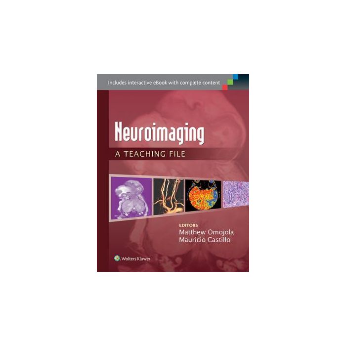
Neuroimaging: A Teaching File [Omojola - LIPPINCOTT Williams and Wilkins]

- ISBN/EAN
- 9781451173284
- Editore
- LIPPINCOTT Williams and Wilkins
- Formato
- Brossura
- Anno
- 2014
- Pagine
- 224
Disponibile
59,00 €
With 100 illustrated cases in print and an eBook containing the 100 print cases plus another200 cases, this volume in the Teaching File Series gives you ready access to actual cases culled from the teaching files of major medical centers. Neuroradiology: A Teaching File covers a wide variety of CNS diseases that are presented as unknowns with coverage of both brain and head and neck cases. Each case features two to four images and follows the Teaching File template: clinical history, findings, differential diagnosis, diagnosis, discussion, questions, key issues for the report, what the clinician needs to know, and answers. It’s the next-best thing to a personal consultation with teaching hospital experts!
Key Features
Clear, concise discussions mimic those performed on a daily basis between residents and faculty members.
Cases are now randomized, to better prepare you for challenges faced in an everyday clinical setting.
Both brain and head and neck cases are highlighted by color perfusion, PET, and functional/MR spectroscopy images.
Common neuroimaging cases are featured, including both normal anatomy and the use of advanced imaging in differential diagnosis.
Collaboration with clinical services and pathology provides an interdisciplinary perspective whenever possible.
Dozens of cases feature full-color images.
New softcover format for the entire series.
Maggiori Informazioni
| Autore | Omojola Matthew; Castillo Mauricio |
|---|---|
| Editore | LIPPINCOTT Williams and Wilkins |
| Anno | 2014 |
| Tipologia | Libro |
| Lingua | Inglese |
| Indice | 1. Treatment related changes 2. Brain stem encephalitis 3. Cerebellopontine angle meningioma 4. Fibrous dysplasia 5. Intralabyrinthine (cochlea) schwannoma (ILS) 6. Enlarged endolymphatic sac syndrome 7. Paget disease 8. Hemangioma of cranial vault 9. Carotid cavernous fistula 10. Orbital and nasofrontal cephalocele 11. Sinus pericranii 12. Hemagioblastoma 13. Posterior fossa ependymoma 14. Hypertrophic olivary degeneration 15. Cross cerebellar atrophy and diaschisis 16. Posterior fossa atypical teratoid rhabdoid tumor 17. Joubert syndrome (JS) 18. Chiari II malformation (CIIM) MR 19. Rhombencephalosynapsis (RES) 20. Creutzfeld Jakob disease (CJD) 21. Alzheimer’s disease (AD) 22 Normal pressure hydrocephalus (NPH) 23. Dural metastasis breast 24. Idiopathic hypertrophic pachymeningitis 25. Neurosarcoidosis 26. Post traumatic CSF collection (pseudomeningocele) 27. Meningitis 28. Epidermoid cyst 29. Subarachnoid hemorrhage (Aneurysmal) 30. Arachnoid cyst 31. Pilocytic astrocytoma 32. Granular cell Astrocytoma 33. Pleomorphic astrocytoma (PXA) 34. Dysembryoplastic neuroepithelial tumor (DNET) 35. Lymphoma 36. Cortical ependymoma 37. Bilateral thalamic glioma 38. Artery of Percheron infarcts 39. Anoxia in term babies 40. Huntington’s disease 41. Post-transplant lymphoproliferative disease (PTLD) 42. Mitochondrial disorder 43. Fahr’s disease 44. Intraventricular simple cyst 45. Choroid plexus papilloma CPP WHO I 46. Intraventricular meningioma 47. Choroid plexus metastasis 48. Central neurocytoma 49. Corpus callosum oligodendroglioma (ODG WHO II) 50. Agenesis of corpus callosum (A CC) 51. Corpus callosum lipoma 52. Corpus callosum tumefactive demyelination (CC TDL) 53. Bilateral middle cerebral artery territory infarctions (MCA infarcts) 54. Cerebral arterial gas embolism (CAGE) 55. Posterior Reversible Encephalopathy Syndrome (PRES) 56. Acute hypertensive basal ganglia hematoma 57. Moyamoya disease (MMD) 58. Thalamic hematoma with spot sign 59. Spontaneous thrombosis of AVM 60. Brain pyogenic abscess with ventriculitis 61. Brain Tuberculomata and abscess 62. Neurocysticercosis vesicular colloidal 63. Mycotic aneurysm left ICA 64. Varicella Zoster virus encephalitis (VZVE) 65. HIV encephalopathy (HIVE) 66. Multiple sclerosis (MS) 67. Tumefactive MS 68. Susac’s syndrome 69. Delayed post hypoxic leukoencephalopathy (DPHL) 70. Paraneoplastic limbic encephalitis (PLE) 71. Methotrexate leukoencephalopathy (MTX LE) 72. Nodular gray matter heterotopia 73. Hydranencephaly 74. Schizencephaly 75. Tuberous sclerosis complex (TSC) 76. Neurofibromatosis type 2 (NF2) 77. Rathke’s cleft cyst 78. Malignant macroadenoma 79. Harmatoma of tuber cinereum 80. Carotid Cavernous fistula 81. Aberrant neurohypophysis 82. Pituitary hemorrhage 83. Vascular supply to the head: The aortic arch variants 84. Azygos anterior cerebral artery (AACA) 85. Cylindrical and Fusiform aneurysms 86. Middle cerebral artery aneurysm with cerebral vasospasm 87. Superior sagittal sinus thrombosis (SSS thrombosis) 88. Vein of Galen aneurysmal malformation (VGAM) 89. Reversible cerebral vasoconstriction syndrome (RCVS) 90. Arteriovenous malformation (AVM) 91. Vasogenic edema 92. Adrenoleukodystrophy 93. Metachromatic leukodystrophy 94. Alexander disease 95. Traumatic intracerebral hematoma (TICH) 96. Traumatic brain injury/Diffuse axonal injury (TBI/DAI) 97. Acute subdural hematoma. (ASDH) 98. Subfalcine herniation (SFH) 99.Traumatic subdural hygroma (TSDG) 100. Chronic calcified subdural hematoma (CCSDH) |
Questo libro è anche in:
